Figure 2.

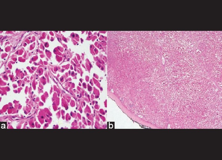
Cells arranged in sheets as well as nests with abundant eosinophilic granular cytoplasm and centrally placed nucleus (a) and Histopathology showing well-encapsulated tumor (b)

Cells arranged in sheets as well as nests with abundant eosinophilic granular cytoplasm and centrally placed nucleus (a) and Histopathology showing well-encapsulated tumor (b)