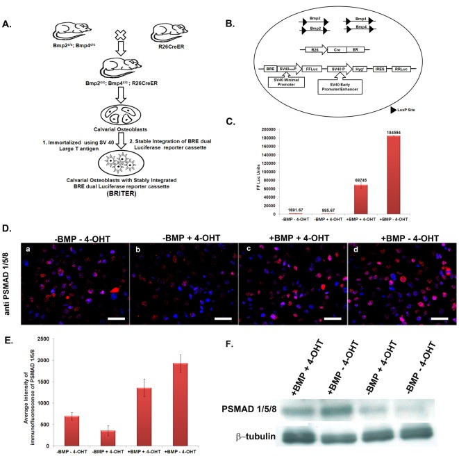
Figure 1. Creation of BRITER Cell Line.
(A) Schematic showing steps involved in the creation of BRITER Cell Line. (B) Schematic showing critical genetic components of BRITER Cell Line. (C) BMP2 dependent FFLuc activity of BRITER Cell Line under four different conditions, namely “−BMP. −4-OHT”, “−BMP, +4-OHT”, “+BMP, +4-OHT” and “+BMP, −4-OHT”. (D) Anti-PSMAD 1/5/8 immunofluorescence in BRITER cell line under four different conditions: (a) −BMP−4-OHT, (b) −BMP+4-OHT, (c) +BMP+4-OHT and (d) +BMP−4-OHT. Scale bar 100 µm. (E) Quantification of Anti-PSMAD 1/5/8 immunofluorescence by Image J. (F) Western blot analysis of BRITER cell extracts cultured under indicated conditions with PSMAD 1/5/8 antibody. β-tubulin antibody has been used as loading control. Data shown are means ± SEM of three independent experiments carried out in triplicates.

