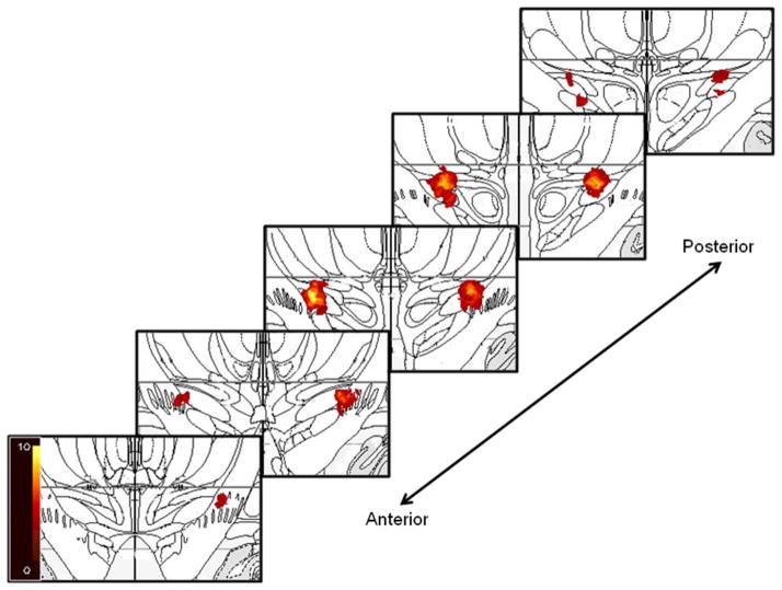
Figure 1.
Location and distribution of the clinically chosen optimal STN DBS electrode contacts superimposed on coronal slices from the Mai atlas 27. For illustration purposes, a 2 mm radius sphere was placed on the center of each active contact as a visual estimate of current spread 28. The scale bar indicates the number of points (participants) at that location.

