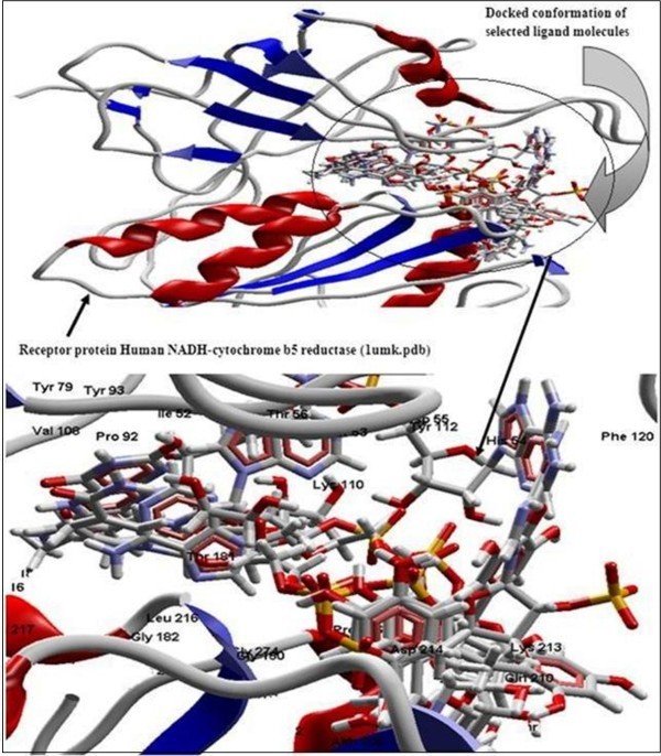
Figure 2.

Secondary structure (cartoon) representation of the active site of receptor human NADH-cytochrome b5 reducatse protein with docked conformation of selected ligand molecules NADPH, beta-NADH, EGCG, quercetin, catechin, epicatechin, resveratrol together with FAD (ligand from crystal structure of 1umk.pdb) using Glide.
