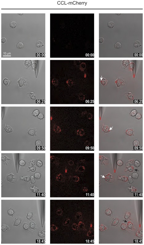
Figure 6. CCL2-mCherry uptake continues after reversal of polarization.
Selected frames from time-lapse video microscopy (Movie S7) of migrating human monocytes recorded at 37°C. The micropipette was dispending 100 nM CCL2-mCherry with constant backpressure. The position of the needle was changed several times during recording. Left panels phase images, middle panels CCL2-mCherry (ex 594 nm/em 620–680 nm), and right overlay. Arrows indicate the localization of internalized CCL2-mCherry in two representative cells upon reverting their axis of polarization. Time stamps are indicated in mm∶ss.

