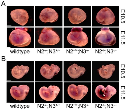
Figure 1. Combined mutations in Notch2 and Notch3 cause defects in vascular development.
Yolk sacs (A) and embryos (B) at E10.5 and E11.5 were dissected and photographed with a stereo microscope. At E10.5, Notch2−/−;Notch3−/− (N2−/−;N3−/−) embryos exhibit a decrease in yolk sac blood vessels, while the embryo proper is relatively normal in appearance. At E11.5, Notch2−/−;Notch3−/− mice show severe vascular defects in both yolk sac and embryo. Yolk sac blood vessels are not visible and extensive hemorrhaging is seen in the embryo (arrowhead). Blood vessels are grossly normal in Notch2−/− (N2−/−;N3+/+) and Notch3−/− (N2+/+;N3−/−) embryos. Scale bar = 1 mm.

