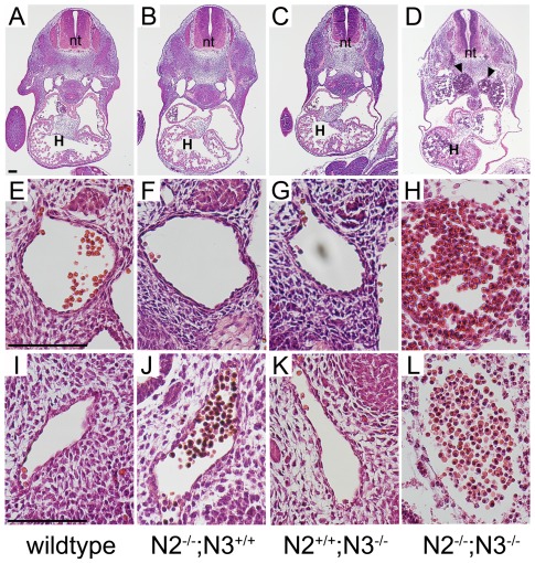
Figure 2. Embryos lacking both Notch2 and Notch3 have disrupted blood vessels.
Hematoxylin and eosin staining of transverse sections of E11.5 embryos through the heart and midsection (A–D), descending aorta (E–H), and caudal aorta (I–L). In the Notch2−/−;Notch3−/− (N2−/−;N3−/−) embryos, the paired dorsal aorta is expanded in size and filled with blood (D, arrowheads). Higher magnification of blood vessels in double mutant embryos show a lack of cells surrounding the lumen (H, L). The overall structure of blood vessels appears relatively normal in the single Notch2−/− (N2−/−;N3+/+) and Notch3−/− (N2+/+;N3−/−) mice. Scale bar = 100 µm. (H) heart, (nt) neural tube.

