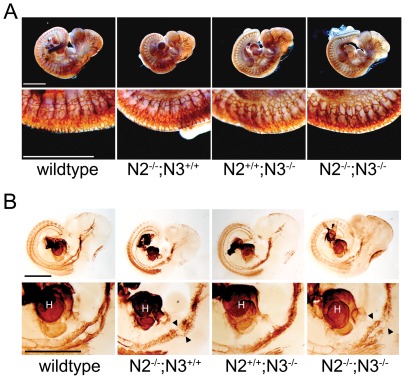
Figure 3. Notch2−/− and Notch2−/−;Notch3−/− embryos develop a normal vascular plexus but have disrupted vascular smooth muscle cells.
Whole-mount embryos at E10.5 were stained for Pecam1 (A) or SMA (B). The vascular plexus is well formed in all mutant embryos with normal vessel patterning seen in large vessels (A, upper panels) and smaller intersomitic vessels (A, lower panels). Whole-mount immunostaining for SMA demonstrates less SMA-positive cells in the dorsal aorta of Notch2−/− (N2−/−;N3+/+) and Notch2−/−;Notch3−/− (N2−/−;N3−/−) (B, lower panels) embryos compared to wildtype and Notch3−/− (N2+/+;N3−/−) embryos. Arrowheads point to paired dorsal aorta. Scale bar = 1 mm. (H) heart.

