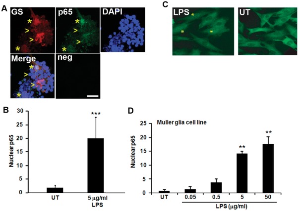
Figure 2. TLR4 is functional in primary Muller glia-photoreceptor co-cultures and a Muller glia cell line.
(A) Nuclear translocation of the downstream TLR4 effector p65 was used as a marker of TLR4 activation. Muller glia-photoreceptor cultures treated with LPS (5 µg/ml) show nuclear localized p65 in Muller glia, as shown by p65 detection in the nuclei of cells costaining with the Muller glia marker glutamine synthetase (GS, asterisks). The arrowheads indicate GS-positive cells that contain cytoplasmic p65, indicating lack of TLR4 activation. Neg, immunostaining negative control. (B) Quantification of cells with p65 signal that overlapped with DAPI stained nuclei in glutamine synthetase-positive Muller glia. Mean ± SD, ***, p<0.001, n = 5. (C–D) MIO-M1 cells treated with LPS also show nuclear p65 (*) compared with untreated cells (UT). Dose-dependent nuclear translocation of p65 by LPS, indicating TLR4 activation in the Muller glia cell line. Mean ± SD, **, p<0.01, n = 5. Scale bar in (A) is 10 µm.

