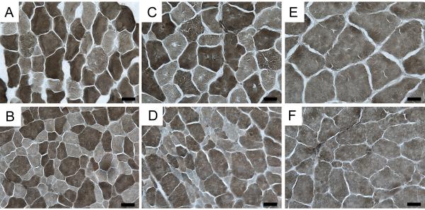
Figure 4.
Photomicrographs of fiber-typed hind limb muscles. TA (A, B), EDL (C, D) and soleus (E, F) from 12 week old SMA affected (B,D F) and control (A,B,C) cats were fiber typed by myofibrillar ATPase reaction at pH 4.6. At this pH, type I (slow twitch) fibers are lightly stained. Both type I and type II (fast twitch) atrophic fibers were observed in affected cats, although the majority of atrophic fibers were Type II. Bars = 20 μm.

