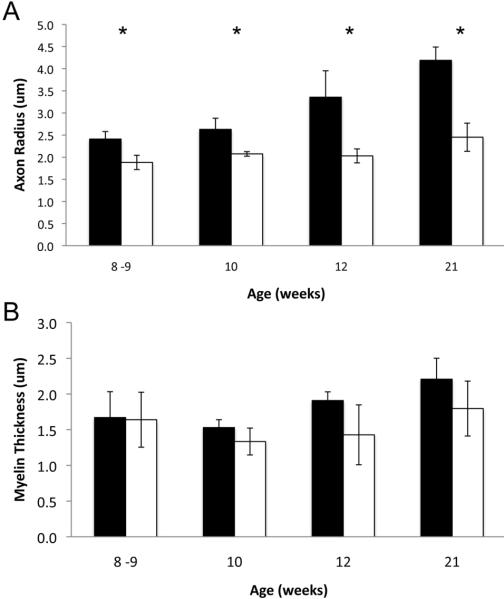
Figure 8.
Average axon radius and myelin thickness. A) Average L5 ventral root axon radius as measured in 100 axons from normal (shaded) and affected (un-shaded) cats. B) Average myelin sheath thickness measured in the same 100 axons (n=3 except at 21 weeks where n=5). Asterisks indicate statistical significance by Student's t-test (p≤0.02).

