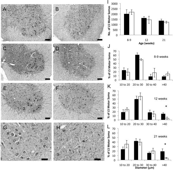
Figure 9.
Cresyl violet stained L5 spinal cord and soma counts and morphometrics. Photomicrographs of L5 anterior horns from normal cats aged 8 wks (A) 12 wks (C) and 21 wks (E) and from age matched affected cats B, D, and F, respectively. Bars=200 μm. G (normal cat, 8 weeks) and H (affected cat, 8 weeks) photomicrographs were taken at a higher magnification (bar = 100 mm) to demonstrate the presence of chromalytic cells (asterisk) and acentric nuclei (arrow) in affected cats. I) Number of motor neuron somas counted in 160 mm of cresyl violet stained L5 spinal cord. Average ± S.D shown. Cell body morphometrics at 8-9 wks (J), 12 wks (K) and 21 wks (L). Averages ± S.D. for normal (shaded) and affected cats (un-shaded) are shown. Asterisks indicate statistical significance by Mann-Whitney U test with p≤0.03.

