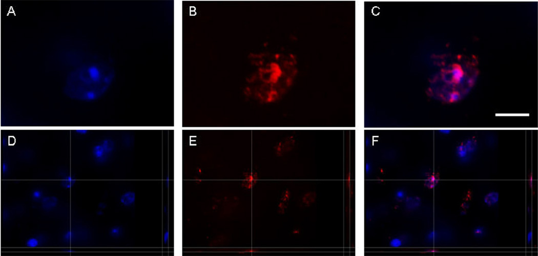
Fig. 2.
5-Methylcytidine (5-mC) and 4′,6-diamidino-2-phenylindole dihydrochloride (DAPI) colocalization. Representative images of a nucleus of a hippocampal cell in a 12-month-old mouse showing fluorescent labeling of DAPI (blue) in (A) and (D), and 5-mC (red) in (B) and (E), at the corresponding location and focal plane, with merged pictures in (C) and (F), showing colocalization (pink). All images represent 1 stack taken with the SI-SD system (see text for more details). Images (D–F) show 3-dimensional reconstructions. Scale bar = 3 µm. Projections and minor corrections in intensity and contrast were made with the Imaris software program (Bitplane AG, Zurich, Switzerland).

