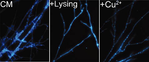
Figure 5.

The abnormal hyphal morphology of the ΔMoswi6 mutant was rescued by the addition of cell wall lysing enzymes or CuSO4. All strains were stained with calcofluor white and fluorescence was mainly distributed on the apex of hyphae and septa. Mycelial morphology was determined under a microscope after the ΔMoswi6 mutants had been grown for 2 days on complete medium (CM) (control) or after the addition of cell wall lysing enzymes or exogenous copper on overlaid microscope slides.
