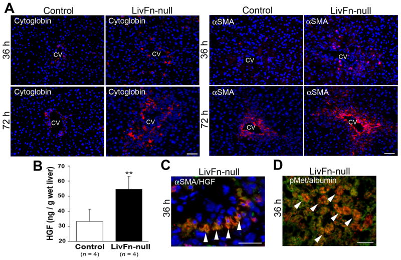
Fig. 5. Fibronectin deficiency significantly upregulates liver HGF levels post injury.
(A) Double immunofluorescence staining for cytoglobin (in red)/DAPI (in blue) and α-SMA (in red)/DAPI (in blue) at 36 and 72hrs post injury. CV, central vein. Bar=50μm.
(B) Hepatic HGF levels at 36hrs post injury. Data are means±S.D. (n=4 for each group). **, P<0.01.
(C) Triple immunofluorescence staining for α-SMA (in red), HGF (in green), and DAPI (in blue) in mutant livers at 36hrs post injury. Arrowheads indicate α-SMA and HGF double-positive activated hepatic stellate cells (orange to yellow color). Bar=25μm.
(D) Double immunofluorescence staining for phospho-Met (pMet, in red) and albumin (in green) in the mutant liver at 36hrs post injury. Arrowheads indicate double-positive hepatocytes (orange to yellow color). Bar=25μm.

