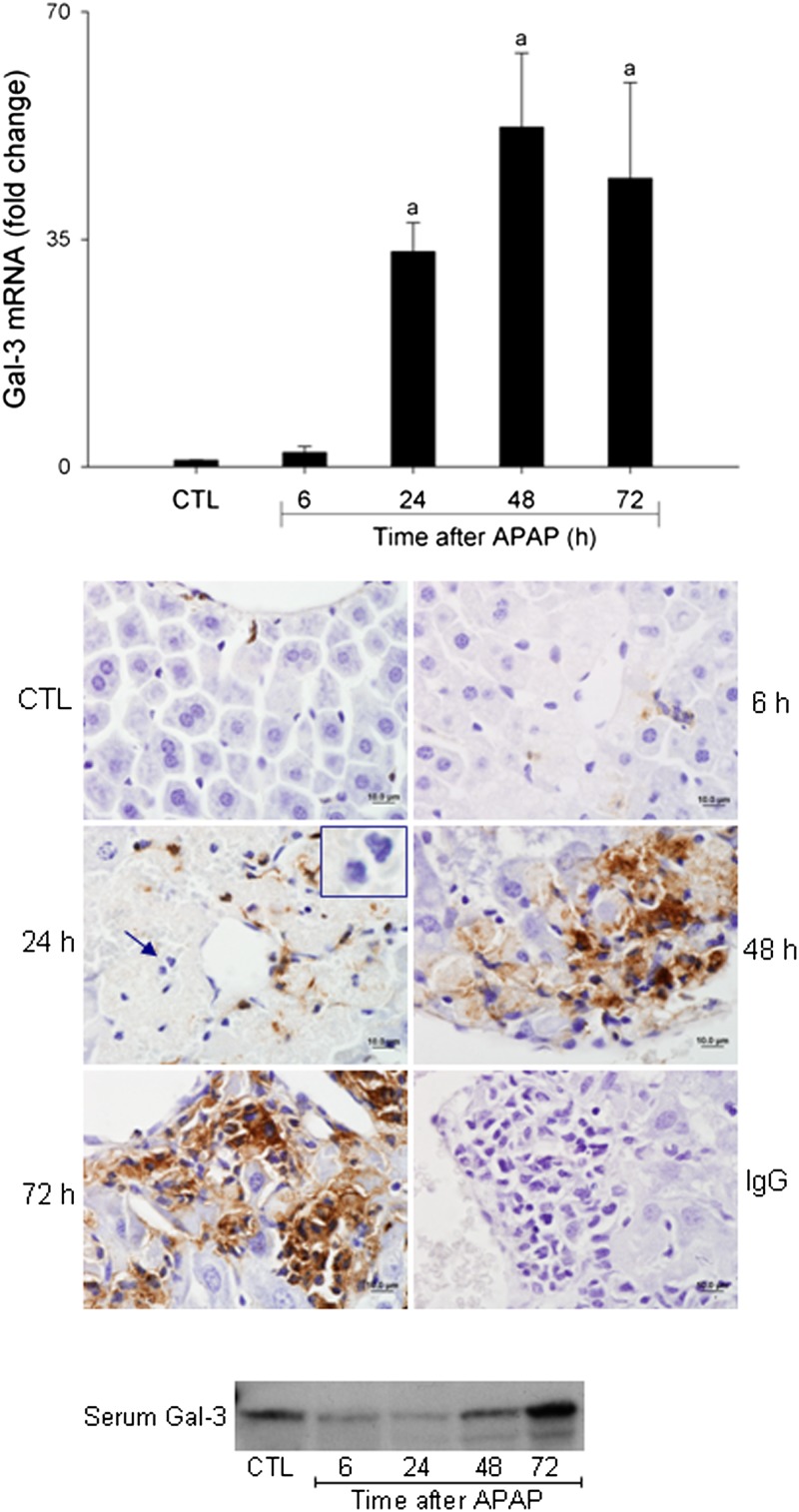
FIG. 1.
Effects of APAP intoxication on Gal-3 expression. Livers were collected 6–72 h after treatment of WT mice with APAP (300 mg/kg, ip) or control (CTL). Upper panel: Gal-3 expression was analyzed by real-time PCR. Data were normalized to 18S rRNA. Each bar represents the mean ± SE (n = 3–10 mice). aSignificantly different (p < 0.05) from CTL. Middle panel: Sections were stained with anti-Gal-3 antibody or IgG control, as described in the Materials and Methods section. One representative section from three independent experiments is shown. Original magnification, ×100. Inset, neutrophils. Lower panel: Serum was collected 6–72 h after treatment of WT mice with APAP or control (CTL). Gal-3 expression was analyzed by Western blotting. One representative blot from five independent experiments is shown.

