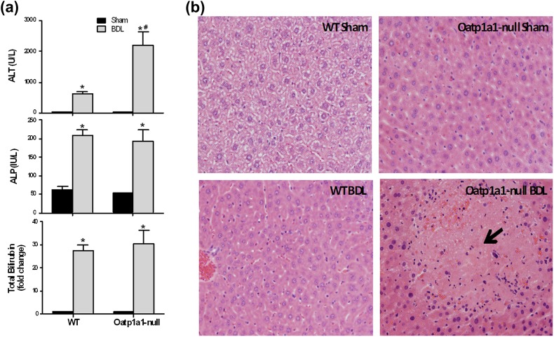
FIG. 1.
Liver toxicity 24 h after BDL in WT and Oatp1a1-null mice. (a) Serum ALT, ALP, and total bilirubin from sham and BDL mice (n = 5 per group) were quantified with analytical kits (Pointe Scientific). All data are expressed as fold change (mean ± SE) for five mice in each group. *Statistically significant difference between sham and BDL groups (p < 0.05). (b) Histological analysis of liver sections from WT and Oatp1a1-null mice 24 h after BDL. Liver sections (5 μm) were stained with hematoxylin-eosin.

