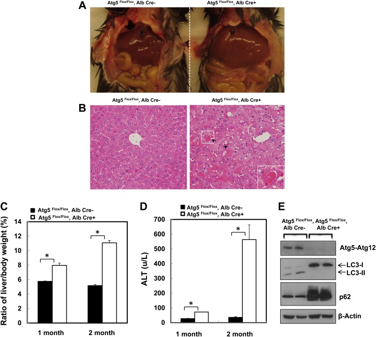
FIG. 1.
Loss of Atg5 in the mouse liver leads to hepatomegaly and liver injury. (A) The gross anatomical views of representative mouse liver from 2-month-old Cre-negative and Cre-positive Atg5 Flox/Flox mouse. (B) Representative photographs of HE staining are presented. Arrows: apoptotic cells. Inserted: enlarged image from the squared area. (C) Ratio of the liver to body weight and (D) serum ALT levels from 1- to 2-month-old Cre-negative and Cre-positive Atg5 Flox/Flox mouse. Data are means ± SE (n = 3). *p < 0.05 Student's t-test. (E) Total liver lysates from 2-month-old Cre-negative and Cre-positive Atg5 Flox/Flox mouse were subjected to Western blot analysis.

