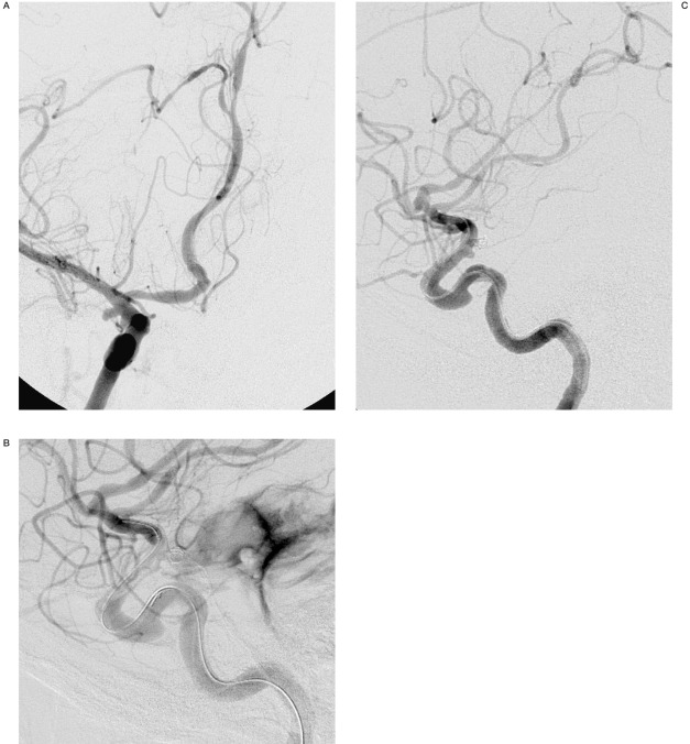
Figure 1.
A) 51-year-old female with subarachnoid hemorrhage. Working projection right internal carotid angiogram demonstrates the anterior choroidal artery aneurysm (arrow) prior to endovascular intervention. B) Right internal carotid angiography following placement of the first coil demonstrates extravasation of contrast into the subarachnoid space (arrow). C) Following inflation of the remodelling balloon across the aneurysm neck for 10-15 seconds, angiography demonstrates cessation of contrast extravasation. The aneurysm is completely occluded (arrow).

