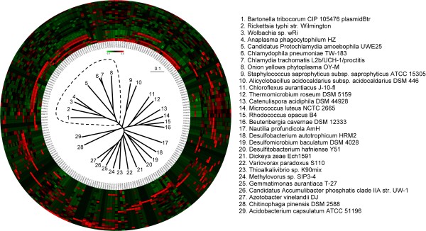
Figure 2.
Probe frequency patterns and clustering result of 29 free/host-living microorganisms. The frequency patterns are represented by the circle; the inner track indicates the probes that can be found in the proteome of these species. These microorganisms, including 8 host-living and 21 free-living species, were clustered according to the similarities of their probe-set frequency patterns by using the CLUSTER 3.0 program, which also generated the classification tree positioned in the center. The host-living and free-living microorganisms are clearly separated. The cluster encircled by the dotted line consists of the 8 host-living species, which correspond to the 8 innermost colored tracks. The positioning of the colored tracks is also determined by the clustering program CLUSTER 3.0.

