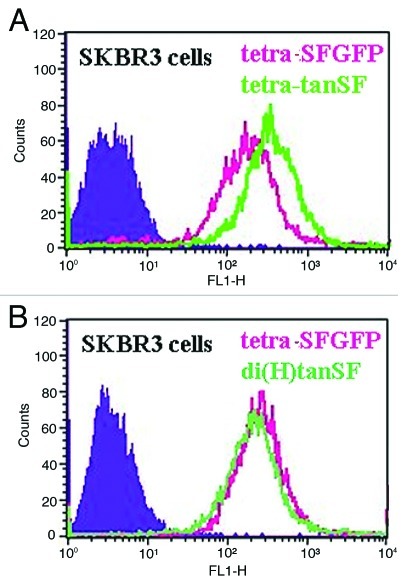
Figure 5. Evaluating the relative contribution of each SFGFP to the overall fluorescence intensity by FACS. SFGFP-Fused antibodies’ ability to stain antigen positive cells was evaluated on human breast adenocarcinoma SKBR3 cells without labeled secondary antibodies. (A) tetraSFGFP compared with tetra-tanSF (B) tetraSF compared with di(H)tanSF. Filled areas, negative control (cell auto fluorescence).

An official website of the United States government
Here's how you know
Official websites use .gov
A
.gov website belongs to an official
government organization in the United States.
Secure .gov websites use HTTPS
A lock (
) or https:// means you've safely
connected to the .gov website. Share sensitive
information only on official, secure websites.
