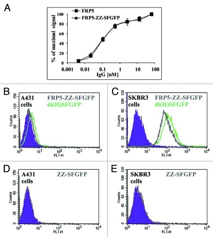
Figure 9. Comparing cytoplasmically folded SFGFP to refolded SFGFP. (A) Binding capacity evaluation by ELISA. Binding of FRP5-ZZ-SFGFP in comparison to the parental Inclonal FRP5 IgG was evaluated on soluble ErbB2 using HRP-conjugated secondary antibodies. Error bars represent standard deviations of triplicates. (B-E) FACS analysis: di(H)SFGFP and FRP5-ZZ-SFGFP ability to stain antigen positive cells was evaluated on human epithelial carcinoma cell line A431 (B) and human breast adenocarcinoma cell line SKBR3 (C). Cells that were incubated with free ZZ-SFGFP alone are shown in D and E as a negative control. Filled areas in all panes; negative control (cells auto fluorescence).

An official website of the United States government
Here's how you know
Official websites use .gov
A
.gov website belongs to an official
government organization in the United States.
Secure .gov websites use HTTPS
A lock (
) or https:// means you've safely
connected to the .gov website. Share sensitive
information only on official, secure websites.
