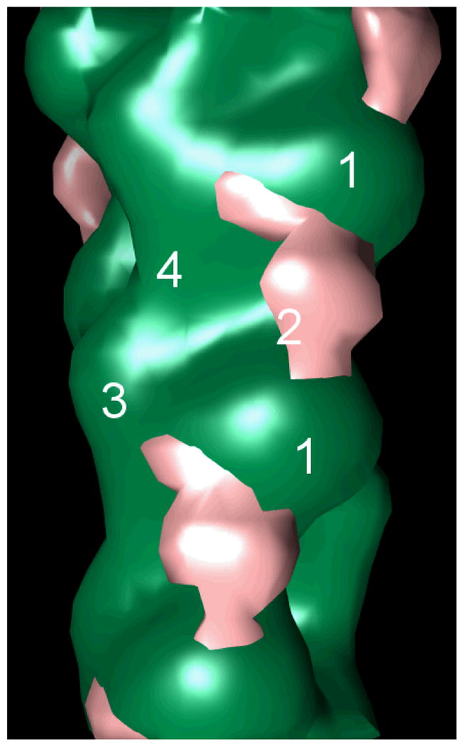
Figure 4. Cryo-electron microscopy of calponin-decorated actin revealed calponin density over actin subdomain 2.

Shown is the difference map of actin (green) with the calponin density (pink) over subdomain 2, bridging subdomains 1 of longitudinally neighboring actin monomers. The four subdomains of one monomer, as well as subdomain 1 of the neighboring monomer, are labeled for spatial reference.
