Figure 1.
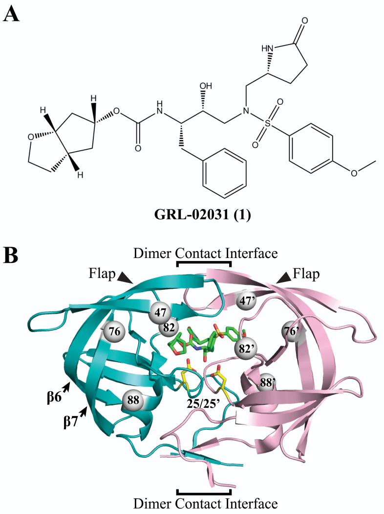
Protease inhibitor 1 and dimer structure of HIV-1 PRWT/1 (PDB ID 3H5B). (A) Chemical structure of compound 1. (B) The backbone of the two subunits is shown in teal and light pink ribbons, the inhibitor 1 is represented by green sticks, and the catalytic residues Asp25/25′ are represented by yellow sticks. The PR structure features of flaps, catalytic pocket, and dimer contact interface are indicated. The arrows indicate the flaps and secondary structure of 6th and 7th β-strands. The dimer interface indicated by the brackets extends from the interacting flap tips through the catalytic triplets to the 4-stranded β-sheet formed by both N- and C-termini. The gray spheres represent the location of mutated residues I47V, L76V, V82A and N88D in both subunits.

