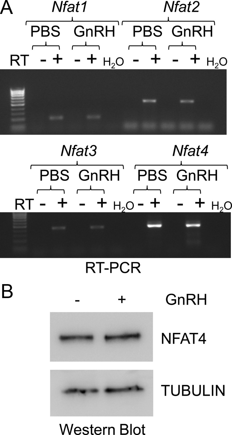
Fig. 4.

LβT2 cells express several Nfat family members. LβT2 cells were serum starved and treated with vehicle (PBS) or GnRH (10 nm) for 1 h. After RNA isolation and reverse transcription of cDNA, semiquantitative PCR was performed using primers specific for Nfat1, Nfat2, Nfat3, and Nfat4 (A). Whole-cell lysates from LβT2 cells were run on a SDS polyacrylamide gel and then immunoblotted for NFAT4 and tubulin, used as a loading control (B). Data shown are representative of three independent experiments. RT, Reverse transcriptase.
