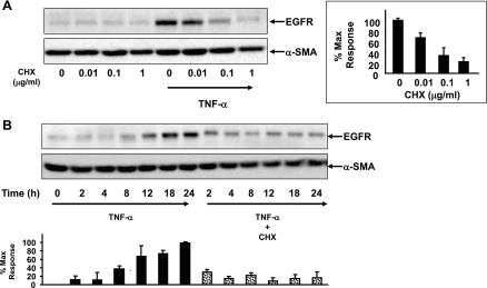
Fig. 2.
Induction of EGFR expression in response to TNF-α requires de novo protein synthesis. A: confluent 18Co cells were washed and equilibrated in serum-free media for 30 min, incubated with cycloheximide at various concentrations (0–1 μg/ml) for 1 h, and then exposed to 10 ng/ml TNF-α for 18 h. Cell lysates were then analyzed by SDS-PAGE and Western blot using an antibody that detects EGFR protein. Results are means ± SE (n ≥ 3) and are expressed as a percentage of the maximum level of EGFR expression, displayed in graphical form at right. Equal protein loading was verified using an antibody that detects α-SMA. B: 18Co cells were pretreated with 1 μg/ml cycloheximide (CHX) for 1 h and then were exposed to 10 ng/ml TNF-α for various times (2, 4, 8, 12, 18, and 24 h, as indicated). Cell lysates were analyzed by SDS-PAGE and Western blot using an antibody that detects EGFR protein. Results are means ± SE (n ≥ 3) and are expressed as a percentage of the maximum level of EGFR expression, displayed in graphical form at bottom. Equal protein loading was verified using an antibody that detects α-SMA.

