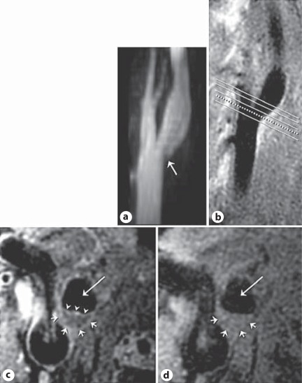
Fig. 1.
MRI images of the right carotid bifurcation in a 46-year-old female without HIV infection or cocaine use. a MR angiogram maximum intensity projection image demonstrates slight indentation into the base of the carotid bulb by an atherosclerotic plaque (arrow). b A long-axis black blood MRI image is used to orient 5 slices through the plaque (lines) along the carotid bulb. c A short-axis postcontrast black blood MRI image oriented through the plaque seen on the long-axis image (dotted line, b) demonstrates the eccentric lesion (short arrows) with an enhancing fibrous cap (arrowheads) separating the lumen (long arrow) from the lipid core (central hypointense tissue). d A precontrast image shows the eccentric plaque (short arrows) and lumen (long arrow) but a relatively inconspicuous core.

