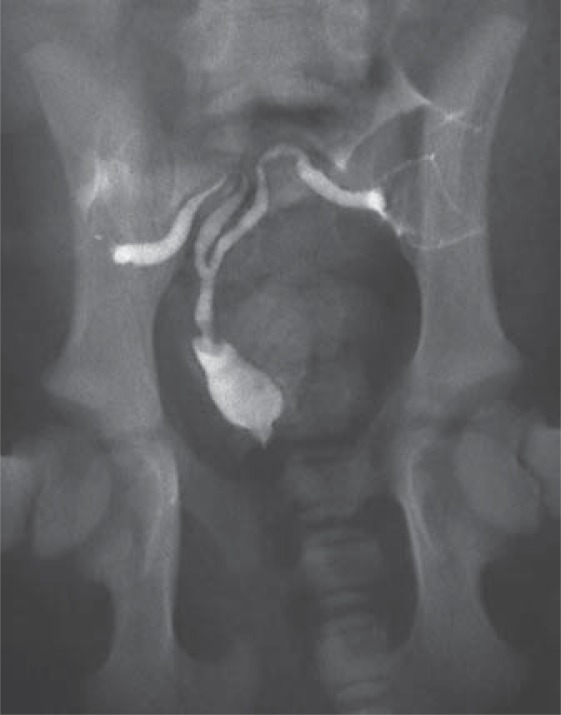
Fig. 7.
XY DSD, PMDS. Ventrodorsal radiograph of a 6-week-old PMDS dog. Contrast dye was injected retrograde via a balloon catheter into the prostatic urethra. Contrast fills the dilated prostatic urethra, the cranial vagina, the uterine body and both uterine horns, confirming a patent connection between the cranial vagina and the prostatic urethra. The prostate parenchyma, which does not contain contrast, surrounds the cranial vagina (radiograph courtesy of Don Schlafer).

