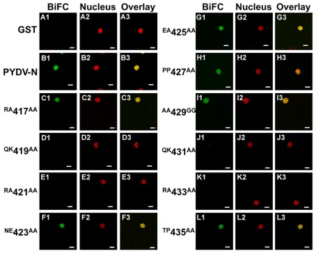
Figure 6.
Confocal micrographs of BiFC assays testing the ability of PYDV-N mutant proteins to associate with the wild-type N protein. Interaction assays were conducted in leaf epidermal cells of transgenic N. benthamiana expressing a fluorescent nuclear marker (Nucleus). Panels 1, 2, and 3 show micrographs of YFP (BiFC), nuclear marker, and the resultant overlay, respectively. Shown here are BiFC assays conducted with wild-type PYDV-N expressed as a fusion to the carboxy-terminal portion of YFP and N mutants expressed as fusions to the amino-terminal fragment of YFP. (A) GST, used as a non-binding control. (B) Wild-type PYDV-N. (C–L) BiFC between PYDV-N and site-directed double mutants of PYDV-N shown in Figure 1. Scale bar = 10 μm.

