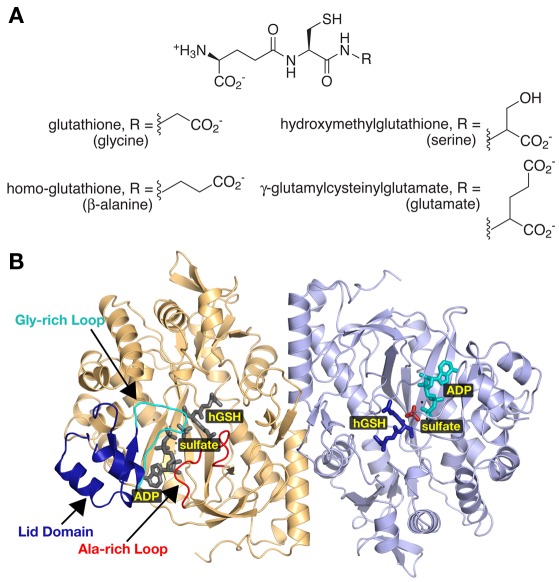
Figure 4.
Diversity in Plant Glutathione Synthetases. (A) The chemical structures of glutathione analogs synthesized by various plants are shown. All share the core γ-glutamylcysteine structure with modifications to the third amino acid position as indicated. (B) Ribbon diagram of the homoglutathione (hGS) dimer (Galant et al., 2009). Monomers are colored gold and blue, respectively. The lid domain (dark blue), glycine-rich loop (cyan), and the alanine-rich loop (red) are highlighted in the gold monomer. Positions of bound ADP, sulfate, and homoglutathione are highlighted in the blue monomer with corresponding ligands colored gray in the gold monomer.

