Figure 5.
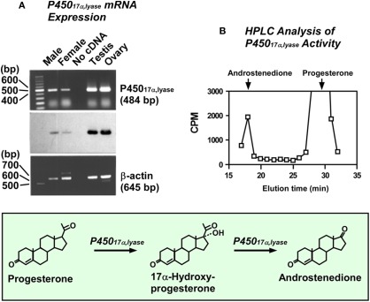
Conversion of progesterone to androstenedione in the quail brain. (A) RT-PCR analysis of cytochrome P45017α,lyase mRNA in the quail brain. Upper panel shows a result of the gel electrophoresis of RT-PCR products for chicken P45017α,lyase, and middle panel shows an identification of the band by Southern hybridization using digoxigenin-labeled oligonucleotide probe for chicken P45017α,lyase. The lane labeled “No cDNA” was performed without template as the negative control. Lower panel shows a result of the RT-PCR for chicken β-actin as the internal control. (B) HPLC analysis of neurosteroids extracted from quail diencephalic slices after 60 min incubation with 3H-progesterone using a reversed-phase column. The column was eluted with a 30-min linear gradient of 40–70% acetonitrile, followed by an isocratic elution of 70% acetonitrile. The ordinate indicates the radioactivity measured in each HPLC fraction. The arrows indicate the elution positions of progesterone and androstenedione. See Matsunaga et al. (2001, 2002) for details.

