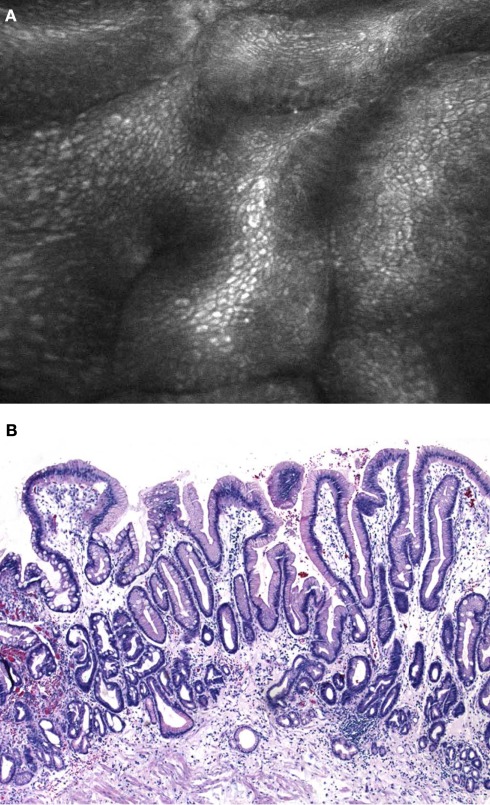
Figure 2.
(A) Confocal laser endomicroscopy using the integrated device (iCLE). Regular shaped columnar lined epithelium with goblet cells (arrows) is clearly visible. (B) Histological image of Barrett’s epithelium with numerous typical goblet cells that replaced the regular squamous epithelium in the distal esophagus (H&E 200×).

