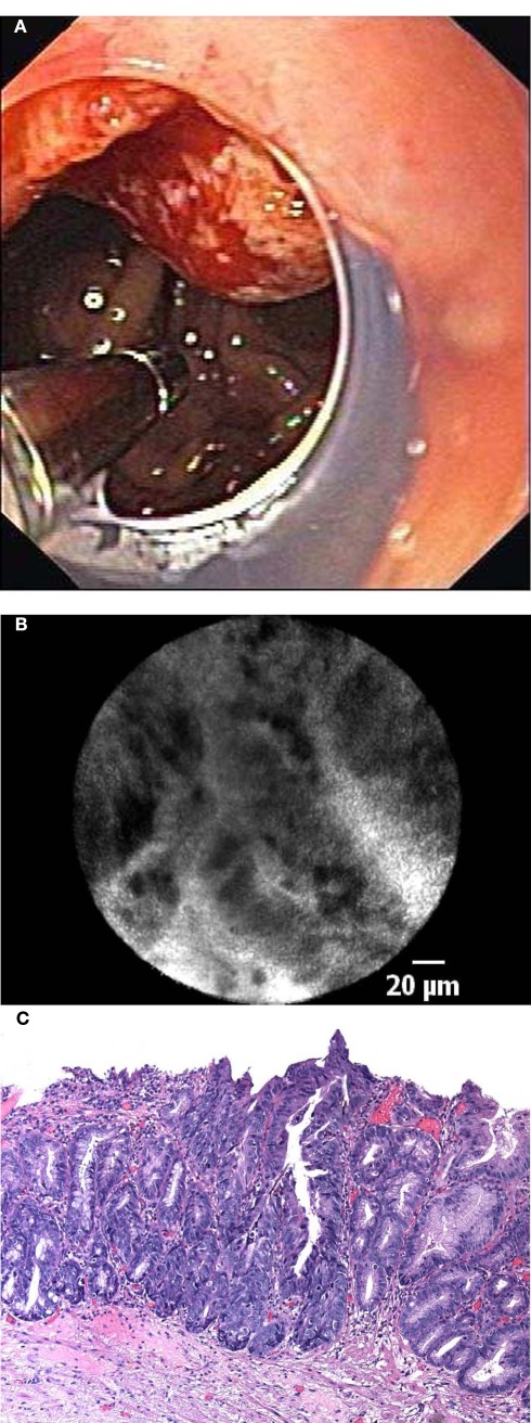
Figure 4.
(A) In vivo endomicroscopy of advanced esophageal adenocarcinoma in patient with long-segment Barrett’s esophagus using the probe-based device (pCLE). Note the clear cap at the distal tip of the endoscope to avoid motion artifacts. (B) Corresponding endomicroscopic appearance of adenocarcinoma in a patient with Barrett’s esophagus. Irregular, dark cells with high contrast to the surrounding tissue indicate advanced neoplasia. (C) Histological image of moderately differentiated adenocarcinoma in a patient with Barrett’s esophagus with loss of regular architecture and superficial erosion.

