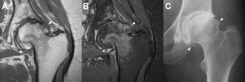
Figure 5.
Marginal osteophytes in a left hip with moderate osteoarthritis as seen with a coronal T1 sequence (A), a coronal proton density fat saturation sequence (B), and plain radiography (C). Marginal osteophytes (arrowheads) are identifiable in all three, but MRI lacks accurate bony definition, while plain radiography lacks useful 3D information. Both can underestimate the extent of osteophytosis. Images courtesy of the Department of Radiology, Addenbrooke’s Hospital, Cambridge.

