Figure 6.
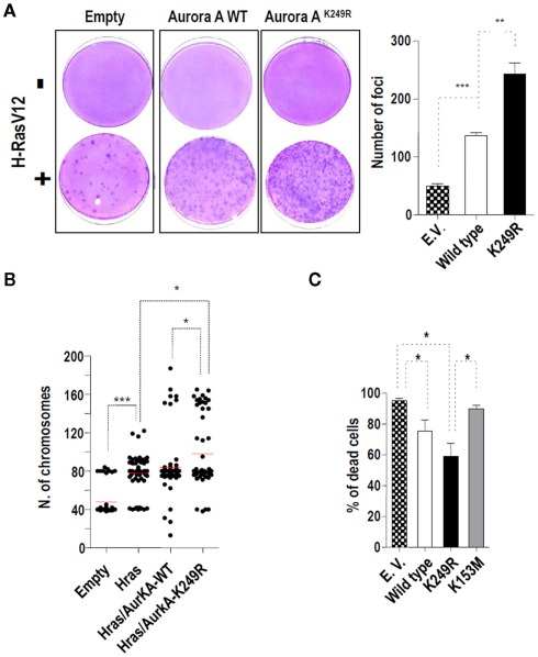
Aurora-A SUMOylation and tumorigenesis. (A) Effects of Aurora-AK249R over-expression on H-RAS-induced transformation in NIH-3T3 cells. Control and Aurora-A expression vectors were co-transfected with an activated G12V–H-RAS vector. Forty-eight hours, post-transfection, the cells were split one to three and kept in culture with 5% calf serum. Two weeks later, dishes were fixed with ethanol and stained with crystal violet. Representative images are shown for each point. A bar graph shows the quantification of the number of foci. (B) Lack of SUMOylation of Aurora-A at lysine 249 increases aneuploidy in H-RAS-transformed mouse fibroblasts. Chromosomes were counted in a minimum of 20 metaphase cells from foci obtained upon transfection with the indicated expression plasmids. Three different foci were analyzed per each transfection type. (C) Lack of SUMOylation increases the taxol chemoresistance induced by Aurora-A over-expression in HeLa cells. HeLa cells stably expressing a control empty vector or EGFP-tagged Aurora-A variants were treated with 150 nM taxol for 48 h. At the end of the treatment, the cells were incubated with propidium iodide (0.02 mg/ml) to determine the number of dead cells (positive for PI). A bar graph shows the quantification for three different clones of each type in one of the two experiments carried out. In all the previous graphs, significant differences are indicated as follows: *p < 0.05, **p < 0.01, and ***p < 0.001.

