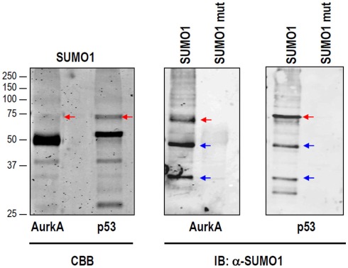
Figure A1.
Non-specific SUMO1-reactive bands are detected upon in vitro SUMOylation. In vitro SUMO1 reactions were performed for Aurora-A and p53 as in Figure 2C. In both cases, the resulting bands were visualized by Coomassie (CBB) staining (left panel). Immunoblotting using an anti-SUMO1 antibody detects a number of common bands (blue arrows) that do not correspond to the expected SUMOylated bands (red arrows).

