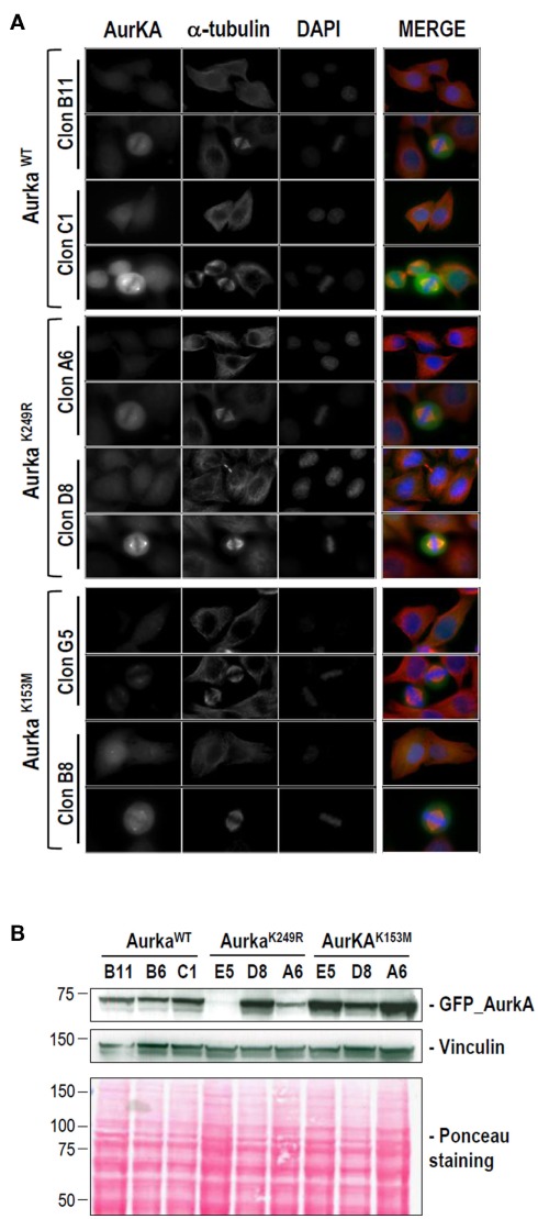
Figure A2.
Stable HeLa cell lines used in this work for the analysis of K249. EGFP-tagged expression vectors were used to transfect wild-type, K249R, and K153M Aurora-A into HeLa cells. G418-resistant, EGFP-positive colonies were expanded and Aurora-A exogenous expression was established by western blotting and immunofluorescence. (A) Representative clones for the expression of high and low levels of EGFP–Aurora-A (green) variants. DNA (blue) and α-tubulin (red) are also shown. Mouse monoclonal anti-α-tubulin (clone DM1A) from SIGMA was used at 1/1000. (B) Detection of EGFP–Aurora-A proteins in stable cell lines by immunoblotting. Three different clones for each Aurora-A variant are shown. Vinculin detection and Ponceau staining are shown as loading controls. The antibodies and dilutions used were as follows: anti-EGFP (Roche, mouse monoclonal; clones 7.1 plus 13.1) at 1/1000 and anti-vinculin (SIGMA mouse monoclonal; clone hVIN-1) at 1/1000.

