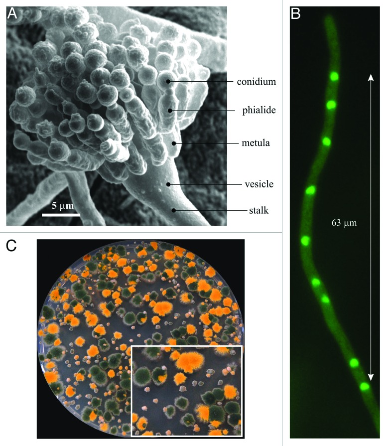
Figure 1.Aspergillus nidulans. (A) Scanning micrograph of a conidiophore with nomenclature of the different specialized cells. (B) A hyphal tip cell. Nuclei were visualized by using a GFP-tagged version of the PacC zinc-finger transcription factor. (C) Progeny of a cross between a rabC+ strain carrying a yA2 “yellow spore” mutation and a rabCΔ strain with wild-type (green) conidiospores. Large colonies are rabC+ progeny whereas small colonies are rabCΔ progeny. As detailed in the text, RabCRab6 plays multiple roles in secretion and therefore its absence results in a conspicuous growth phenotype. The inset displays a sector of the Petri dish at double magnification.

An official website of the United States government
Here's how you know
Official websites use .gov
A
.gov website belongs to an official
government organization in the United States.
Secure .gov websites use HTTPS
A lock (
) or https:// means you've safely
connected to the .gov website. Share sensitive
information only on official, secure websites.
