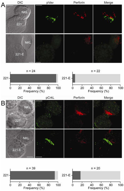
Figure 4. Phosphorylated Crk Accumulates at Inhibitory Synapses between NKL and 221-E Cells.
NKL cells were conjugated with the HLA class I-negative human cell line 721.22l (221), and 221 cells expressing HLA-E (221-E) for 60 min. DIC images are shown on the Left. Confocal microscope z-series were obtained and projected serial confocal sections are shown. (A) Fixed and permeabilized cells were incubated with Abs to pY174-Vav1 followed by Alexa Fluor 488-conjugated secondary Abs (Green). Perforin was stained with a primary Ab followed by Alexa Fluor 647-conjugated secondary mAb (Red). Merged overlays are on the Right. For conjugates with 221 cells, the dark shaded bar indicates the frequency of cells displaying polarization of perforin and pVav-1. For conjugates with 221-E, the dark shaded bar indicates cells displaying polarization of p-Vav1, but not perforin; the light shaded bar indicates cells with neither perforin nor p-Vav1 polarization. (B) Fixed and permeabilized cells were incubated with Abs to pY207-CrkL followed by Alexa Fluor 488-conjugated secondary Abs (Green). Perforin was stained as in (A). For 221 conjugates, the dark shaded bar indicates cells displaying polarization of perforin and no p-CrkL. For 221-E conjugates, the dark shaded bar indicates cells displaying no perforin polarization and no p-CrkL; the light shaded bar indicates cells displaying polarization of p-CrkL, but not perforin. Scale bars are 2.0 μm. The images are representative of 20 cells in two independent experiments.

