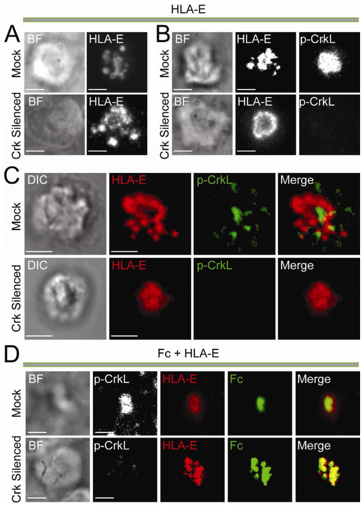
Figure 6. HLA-E Promotes Crk-Independent Central Clustering of Fc.
(A–C) Images of NK cells transfected with control siRNA (Mock) and Crk siRNAs (Crk silenced) and incubated on lipid bilayers carrying HLA-E-Alexa Fluor 568. Brightfield (BF) images are shown on the Left. (A) TIRF images of live NK cells at 30 min. (B) TIRF images of NK cells fixed after 30 min and stained with pY207-CrkL primary Ab followed by Alexa Fluor-488-conjugated secondary Ab. (C) 3D confocal imaging of NK cells fixed ~60 min after addition to the lipid bilayer. Staining for pY207-CrkL (Green) was as in (B). (D) TIRF images of NK cells fixed ~50 min after addition to a lipid bilayer carrying Fc-Alexa Fluor 488 (Green) and HLA-E-Alexa Fluor 568 (Red), and stained for pY207-CrkL with primary Ab followed by Alexa Fluor 647-conjugated secondary Ab (White). Treatment with siRNAs was as in (A–C). Scale bars are 3.0 μm. The images are representative of at least 100 cells in three independent experiments.

