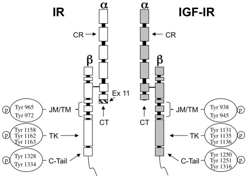
Figure 1.
Structure of IR and IGF-IR and autophosphorylation sites. The ligand binding sites of both receptors are predominantly located at a cysteine-rich region (CR) in the extracellular α-subunit. The homology between IR and IGF-IR in this region ranges 45–65%. The CT peptide in the α-subunit contributes to the binding properties of both receptors. In IR the hatched fragment on the bottom of the CT region is encoded by exon 11 and is present in IR-B isoform but not in IR-A. The tyrosine kinase domain (TK) in the β-subunit is highly conserved showing 85% of similarity between both receptors. The most divergent region is the C-terminal domain (C-tail). JM, juxtamembrane domain; TM, trans-membrane domain.

