
Figure 8.
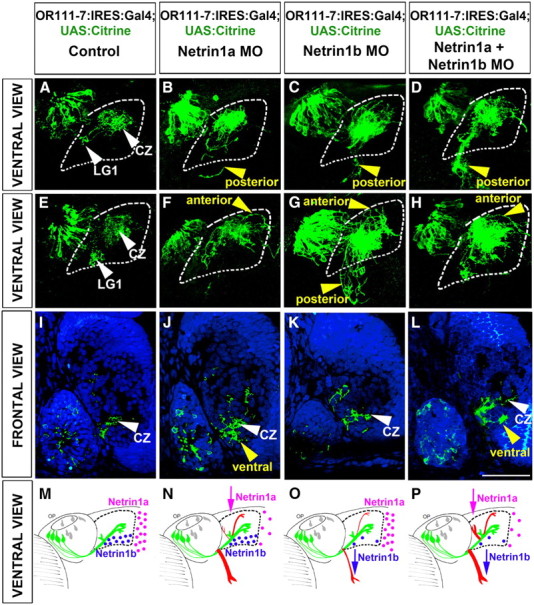
Netrin1a and netrin1b contribute to the proper pathfinding of OR111-7-transgenic axons into and within the olfactory bulb. A–H, Maximum intensity projections of serial confocal optical sections of 3-d-old larvae (ventral view). Anterior is to the top and the midline is to the right. The olfactory bulb is outlined with dashed lines. M–P, Schematics showing control OR111-7:IRES:Gal4; UAS:Citrine projections (green) and mistargeted axons (red) observed upon inhibiting netrin1a, netrin1b or netrin1a and netrin1b together. The thickness of the red lines corresponds roughly to the penetrance of the indicated phenotypes. Netrin1a mRNA (pink dots) is expressed at the telencephalic midline and netrin1b (blue dots) is expressed in the ventral bulb. A, E, M, OR111-7:IRES:Gal4; UAS:Citrine axons (green) project to the central zone (CZ, white arrowhead) and LG1. B–D, N–P, Some OR111-7-transgenic axons misproject posteriorly (posterior, yellow arrowheads) rather than entering the olfactory bulb when Netrin1a, Netrin1b, or both Netrin1a and Netrin1b levels are reduced. F–H, N–P, OR111-7:IRES:Gal4; UAS:Citrine axons also inappropriately stray anteriorly (anterior, yellow arrowheads) in netrin1a, netrin1b, or netrin1 and netrin1b double morphants. I–L, Single confocal optical sections of 3-d-old zebrafish larvae (frontal view). Dorsal is toward the top of the image. Propidium iodide (blue) labels olfactory bulb cells and allows identification of distinct protoglomeruli. I, OR111-7 transgene-expressing axons are observed in the central zone (CZ, white arrowhead) in uninjected Control larvae. J, L, Reducing Netrin1a levels or Netrin1a and Netrin1b levels together causes ventral mistargeting of OR111-7 transgene-expressing axons (ventral, yellow arrowhead, schematized in Fig. 5R,T). Scale bar (in L): A–L, 50 μm.