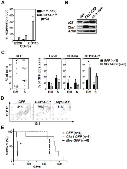
Figure 4. Ubiquitous Cks1 overexpression in the bone marrow is not sufficient to induce myeloproliferative disease or leukemia.
4×10⁶ GFP-positive cells were transplanted with an infection rate of 30%. A, Cks1 mRNA levels in the indicated bone marrow subsets of transplanted mice were analyzed by realtime PCR at day 160 after transplantation. Shown is the relative expression of Cks1 as compared to the B cell expression of the GFP mice as a control sample set at 1. GFP group: n = 2; Cks1-GFP group: n = 3. The bars represent the mean relative expression ± standard deviation. realtime PCR was performed in duplicates. B, Immunoblot analysis of the bone marrow of transplanted mice on the day of indisposition and sacrifice. C, The left panel shows the GFP positivity of the bone marrow (BM) and spleen (S). The three right panels show the percentages of B-, T- and myeloid cells. D, Representative flow cytometric dot blot images of the myeloid cell compartment of GFP, Cks1-GFP and Myc-GFP animals on the day of their sacrifice. E, Survival analysis of lethally irradiated recipient mice that were transplanted with GFP, Cks1-GFP or Myc-GFP infected bone marrow. None of the GFP or Cks1-GFP mice died of leukemia, while all Myc-GFP mice succumbed to AML. Median survival: GFP group, 469 days; Cks1-GFP group, 541.5 days; Myc-GFP group, 52 days. Only the survival of Myc-GFP group is significantly different from the other groups.

