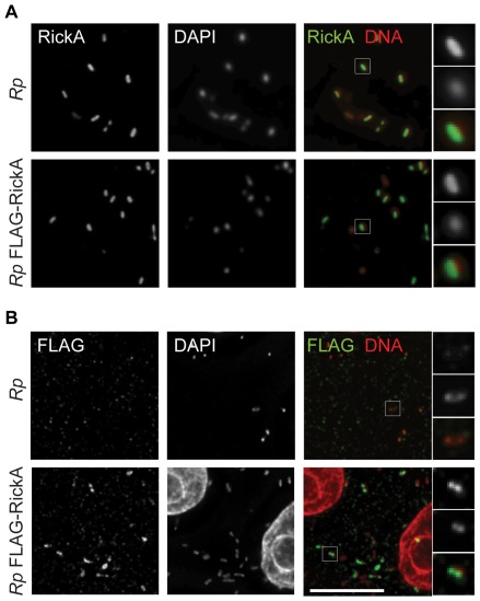
Figure 5. Detection of FLAG-RickA in bacteria.
R. parkeri strains that were not transformed (Rp) or transformed with pMW1650-FLAG-RickA (Rp FLAG-RickA) were used to infect Vero cells and then (A) labeled by immunofluorescence with anti-RickA antibody and stained for DNA with DAPI, or (B) labeled by immunofluorescence with anti-FLAG antibody and stained for DNA with DAPI. In the merged images, RickA or FLAG are labeled in green, and DNA in red. Scale bar 10 µm. Higher magnification images of individual bacteria (highlighted in boxes in the lower magnification images) are shown on the right.

