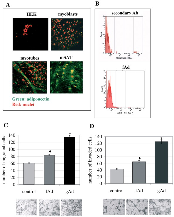
Figure 4. mSAT are a source of autocrine fAd.
A) Analysis of adiponectin expression in Human Embryonic Kidney (HEK) cells, C2C12 murine myoblasts, four days differentiated C2C12 and mSAT by confocal microscopy. Cells were seeded on cover slip, fixed and then treated with anti-adiponectin antibodies and with propidium iodide to label the nuclei. The visualization of adiponectin was performed using Alexia Fluor 488-coniugated secondary antibodies. The images are representative of four independent experiments. B) Analysis of adiponectin expression by citofluorimetric analysis. Cells were treated as in A) and then analyzed using a FACSCanto cytofluorimeter. C) Migration of mSAT induced by fAd and gAd. mSAT were serum deprived overnight and then seeded in the upper chamber of the Boyden assay in free-serum medium supplemented with fAd (1 µg/ml) or gAd (1 µg/ml). Four days differentiated C2C12 myotubes were cultured in the lower chamber and used as chemo-attractant. D) The invasiveness assay was performed as described in C, except for the presence of a thin layer of Matrigel in the upper chamber. Images are representative of four independent experiments and the mean of migrated cells in six independent experiments was shown in the bar graphs. *p< 0,002 and ♦p<0,01 vs control.

