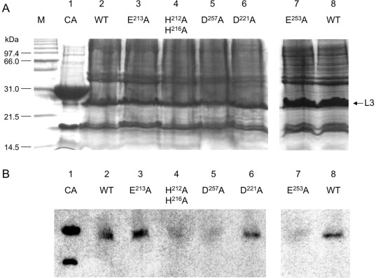
Figure 4. Coomassie Blue-stained SDS-PAGE of partially purified inclusion bodies of wild-type and L3 loop mutants (A) and Zinc blot analysis (B).
In A and B: 1: bovine carbonic anhydrase (AC) (∼30 kDa; 5 µg), was used as positive control and 2 to 6 are, respectively, wild type (WT), E213A, H212A/H216A, D257 A, D221 A and E253 A L3 loop mutants. For each gel 40 µg of inclusion bodies were loaded. The fluorographies were exposed at -70°C for 72 hours. M: molecular weight marker. The arrow indicates L3 loops.

