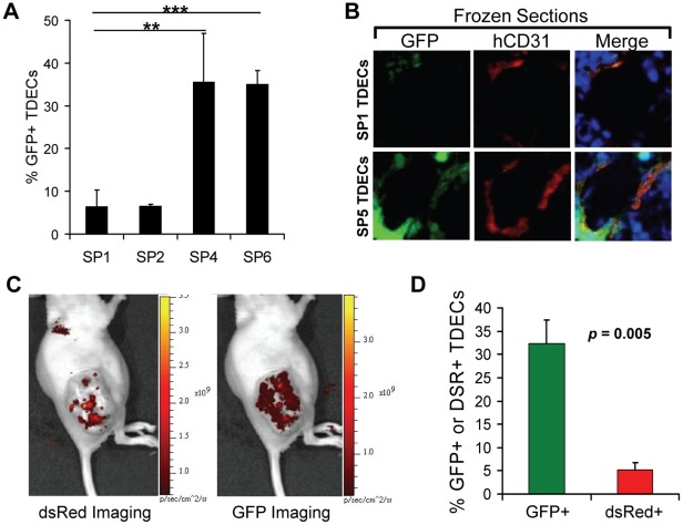
Figure 1. Increased tumor re-populating ability of serially-passaged TDECs.
(A) Human and murine TAECs were isolated from initial H460 lung cancer xenografts by CD31 immuno-affinity purification [16]. The latter were then eliminated by propagating the cells in G-418 and the resultant GFP+, G418-resistant human TDECs were then serially passaged in vivo with a 20-fold excess of non-GFP-tagged H460 tumor cells. At the indicated passage numbers, the percent of GFP+ TDECs was determined 1–2 days after isolation and before the addition of G-418. The results shown represent the average values obtained from 3–5 fields (±SEM) with a total of 738, 761, 128, and 296 cells counted for SP1, SP2, SP4, and SP6, respectively. The statistical analysis was performed using a two-tailed Student’s t test (**, p<0.01; ***, p<0.0001). (B) Photomicrographs of typical blood vessels from H460 tumor xenografts established as described in A for serial passages SP1 and SP5. Frozen sections were visualized for GFP and stained for human-specific CD31 (hCD31) (red) and DAPI (blue). Images were taken at 20× magnification. (C) Live animal imaging of tumor xenografts arising in a representative mouse co-inoculated with an equal number of dsRed-tagged SP0 TDECs and GFP-tagged SP6 TDECs plus a 20-fold excess of untagged H460 tumor cells. (D) Quantification of the percent red SP1 versus the percent green SP7 TDECs isolated from three tumors. The graph represents the mean values (±SEM) with the p value determined using a one-tailed Student’s t test.

