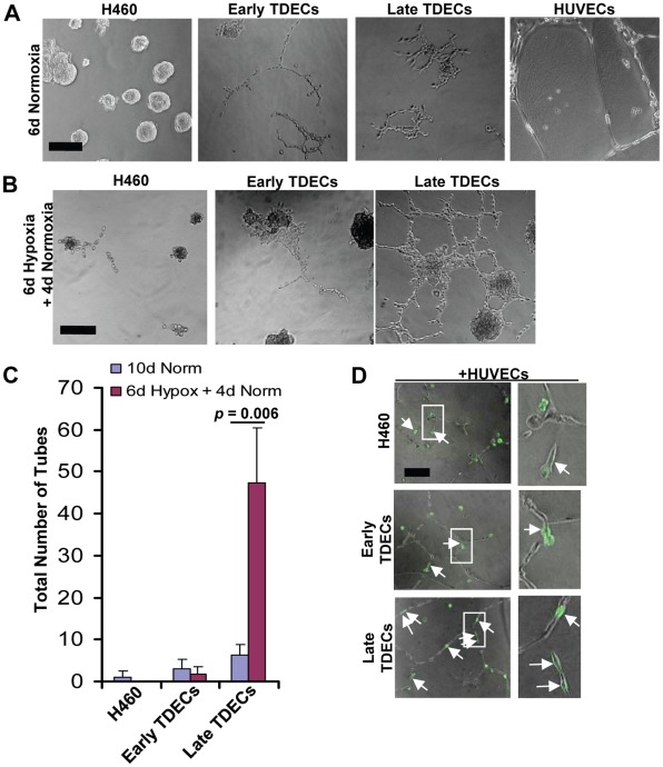
Figure 3. Serially-passaged TDECs demonstrate increased tube formation.
(A) H460 tumor cells, early and late passage TDECs, and HUVECs were plated in Matrigel as previously described [16] and incubated under standard normoxic conditions for six days. Typical light microscopic fields are shown. All photos are shown at identical magnifications with the black bar representing 100 µm. (B) The experiment from A was repeated except that cultures were incubated for six days under moderately hypoxic conditions (1% O2) followed by an additional four day recovery period under normoxic conditions. (C) The total number of tubes from B as well as those from parallel plates of cells cultured 10 d in normoxia (control) were quantified. The plot depicts the mean values (±SEM) from three independent experiments. The p value was determined for only the late TDECs using a one-tailed Student’s t test. (D) GFP-tagged H460 tumor cells or early or late TDECs (1,000 cells) incubated alone or mixed with non-GFP-tagged HUVECs (10,000 cells). The cells were then cultured for two days under normoxic conditions in a standard Matrigel-based tube assay. Brightfield and UV fluorescence photographs were taken and the merged images shown. In the first two cases, the white arrows indicate GFP+ tumor cells or early-passage TDECs that lie adjacent to but have not incorporated into small groups of HUVECs. Arrows in the bottom two panels indicate late passage TDECs that have incorporated into HUVEC tubes. Note that when GFP-tagged H460 tumor cells and early and late GFP+ TDECs were plated without HUVECs, they remained rounded after 2 days of culture on Matrigel (see Figure S1). All photos are shown at identical magnifications with the black bar representing 100 µm. Enlargements of representative views (areas in white rectangles) are also shown. Original images in panels A, B, and D were taken at 10× magnification.

