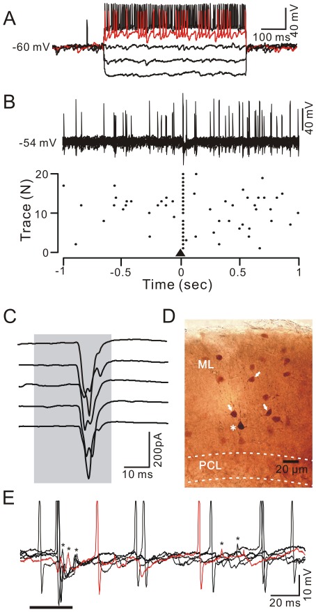
Figure 2. Air-puff stimulation of the ipsilateral whisker pad evoked spike firing in stellate-type MLIs.
A, Whole-cell patch-clamp recording from a stellate-type MLI in response to hyperpolarizing pulses (−100 pA) followed by a series of depolarizing current pulses (+50 pA/step). B, Under current clamp (I = 0), superposition of 20 consecutive traces (upper) and a raster plot of spike firing (lower) showing the stellate-type MLI response to air-puff stimulation (black triangle, 30 ms). Time point 0 denotes the onset of the air-puff stimulation. C, Under voltage-clamp (Vhold = −70 mV), five sequential traces showing air-puff stimulation (bar, 30 ms)-evoked EPSCs in the stellate-type MLI. D, A photomicrograph depicting the morphology of the stellate-type MLI (asterisk) filled with biocytin and dye-coupled to several other MLI s (arrows). E, Under current-clamp conditions (n = 0), example traces (n = 5) showing the air-puff stimulation-evoked spike firing or spikelet discharge in a stellate-type MLI. Spikelet discharges are indicated by asterisks.

