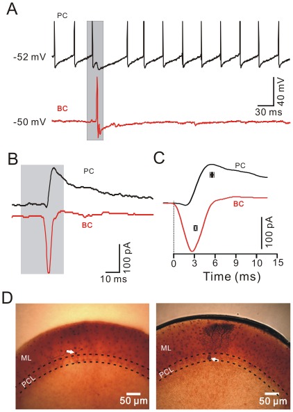
Figure 4. Comparison of the air-puff stimulation-evoked responses in a basket.
-type MLI and a PC in the same mouse cerebellar Crus II. A, Under current-clamp (I = 0) conditions, air-puff stimulation (grey shadow) evoked spike firing in a basket-type MLI (lower), and an IPSP with a pause in spike firing in a PC (upper), in the same mouse cerebellar Crus II. B, Under voltage-clamp (Vhold = −70 mV), air-puff stimulation (grey shadow) evoked fast EPSCs in the basket-type MLI (lower) and IPSCs in the PC (upper). C, Enlarged current traces from (B) and the mean values (± SEM) of the time to peak for the current traces evoked by air-puff stimulation in the PC (black; n = 5) and the basket-type MLI (red; n = 5). D, Consecutive photomicrographs showing the basket-type MLI (white arrow; left) and the PC (white arrow; right) filled with biocytin. The two recorded cells were apart from ∼150 µm in coronal plane. PCL, PC layer; ML, molecular layer.

